|
|
 |
Sinogram Restoration for Ultra-low-dose X-ray Multi-slice Helical CT by Nonparametric RegressionThe objective of this study is to decrease noise levels and streak artifacts in multi-slice, ultra low-dose helical CT images and find an alternative technique to reconstruct images from a multi-slice low-dose helical CT sinogram. This could ultimately lead to substantial dose reduction in routine CT studies.
Recently, increasing attention has been paid to the annual screening of smokers and former smokers for early-stage lung cancer detection. However, because a larger population will be involved in x-ray CT lung cancer screening exams, radiation exposure becomes an important concern due to its association with radiation-induced lung cancer. Reducing radiation exposure of CT, i.e. lower tube current, will inevitably increase noise levels in the sinogram as well as degrade the quality of reconstructed CT images. One manifestation of this are streak artifacts near the apices of the lung. In addition, researchers have documented that the radiation exposure in a current generation multi-slice helical CT could be up to 4 times higher than a conventional non-helical scanner in many clinical applications. Multi-slice helical low-dose CT images induce non-uniform noise with noise levels more than 10%-40% of that associated with conventional CT images.
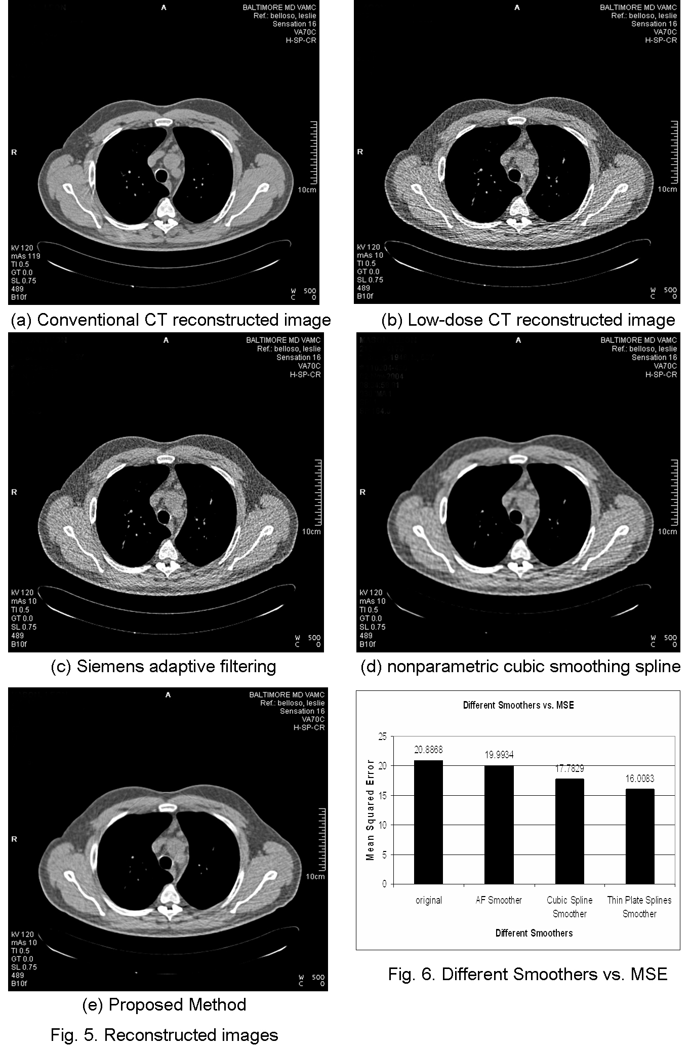
Previous researchers have proved that the noise model in low-dose CT projection profiles after system calibration follow an Gaussian distribution and that the roughness penalty based nonparametric regression has been one of the most optimal choices in low-dose CT projection profile smoothing. In this research, the aforementioned work has been extended into 2D low-dose CT projection frames. A natural generalization of smoothing splines, known as thin plate splines, is applied to the 2D sinogram.
In this research, the noise in each low-dose CT projection was reduced by leveraging the information contained not only within each single projection, but also among nearby projections. The results showed the effectiveness of the proposed approach, which not only suppressed the noise in the original ultra-low-dose sinogram to a minimum, but also preserved valid high frequency information, such as boundaries, etc. The comparison results favor the proposed method, which yields a reconstructed CT image with better contrast and less noise compared to the conventional 1D based cubic spline smoothing approach. |
| [Home] [Biography] [Research] [Teaching] [Industry Exp.] [Lab. Personnel] [Collaborators] [Publications] [Links] |

