|
|
 |
Non-invasive Imaging Techniques as a Quantitative Analysis of Skin Damage Due to Ionizing Radiation
The response of the skin to ionizing radiation has important implications for both the treatment of malignant disease by radiation and for radiological protection. After undergoing radiation therapy as cancer treatment, a patientís skin responds in many ways; one of the most debilitating is the formation of fibrosis, or thick rough skin. Radiation damage, including fibrosis, can be permanent, but its onset and chronic extension have been poorly understood. The objective of this research was to develop a means of optically and non-invasively quantifying radiation damage in mouse skin. Some of the effects of radiation have been documented in pathological, biochemical, and cell culture studies. These experiments have shown radiation-induced changes in collagen production, vascularization, protein levels, and the structure and cell populations of the epidermis and dermis layers of the skin.
This study tested the ability of two non-invasive techniques, thermography and near-infrared multi-spectral imaging, to quantitatively assess the response of mouse skin to a single dose of X-ray irradiation. Thermal images from an 8-12 micron thermal camera were recorded after a cold stimulation to see the thermal recovery of the skin. The irradiated areas showed a significantly faster thermal recovery than the non-irradiated areas two weeks after radiation (p < 0.05).
The NIR multi-spectral imager obtained images at six specially selected wavelengths between 700 and 1000 nm. Two-layer model-based diffuse reflectance spectroscopy monitored changes in blood oxygen saturation and blood volume. Blood oxygen fractions were significantly lower after radiation (p < 0.05). Blood volume changed in six of seven irradiated mice one week after radiation. The non-invasive imaging techniques were successful in quantitatively analyzing the response of the skin to a single dose of irradiation.
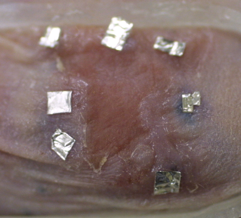
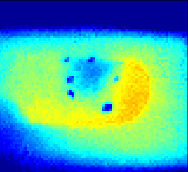
32 Gy mouse (#2) two weeks after radiation in (a) Digital image with aluminum foil pieces covering the India ink tattoos. Red area inside tattoos is irradiated area showing desquamation; and (b) Static thermal image before cold stimulation showing affected areas to be colder in the image.
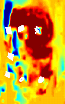
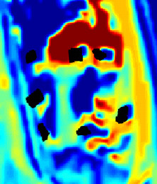
Oxygen blood saturation images of 32 Gy (#2) mouse (a) at day 0 before radiation; and (b) two weeks after radiation was applied. High oxygen saturation inside the radiated area before radiation was no longer present two weeks after radiation. |
| [Home] [Biography] [Research] [Teaching] [Industry Exp.] [Lab. Personnel] [Collaborators] [Publications] [Links] |


