|
|
 |
Using Multi-Modality Imaging Techniques to Assess Vascularity in AIDS-Related Kaposiís Sarcoma
The visible inspection and palpation of skin lesions have long been used to assess the course of cutaneous disease in patients with Kaposiís sarcoma (KS). However, reliable assessment requires a highly trained evaluator and evaluations made by different observers or by the same observer at different times can be inconsistent. Quantitative instrumental methods offer a potentially more objective means of assessing skin health to supplement the visual clinical observations. Moreover, such approaches can be used to provide early markers for tumor responses and to learn about the pathophysiology of the disease and its changes in response to treatment.
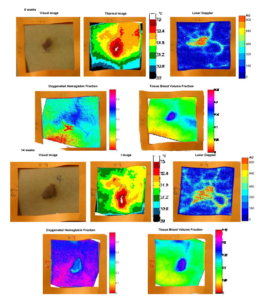
This study analyzed AIDS-related KS lesions with three quantitative non-invasive techniques: thermography, laser Doppler imaging and near-infrared (NIR) spectroscopy. Optical spectroscopy is most related to visual assessment. With Lawrence Livermore National Laboratory, we designed a spectral imager with six wavelengths (700, 750, 800, 850, 900, and 1000 nm). Local variations in melanin, oxy-hemoglobin (HbO2) and blood volume were found.
The spectroscopic data was combined with 8-12 micron infrared and 780 nm laser Doppler images obtained from the same area of the patient. Thermography graphically depicts temperature gradients and has been used to study biological thermoregulatory abnormalities that directly or indirectly influence skin temperature. Thermography provides an integrated thermal signature that combines deep and surface sources and in general can be related to increased blood flow associated with increased metabolic activity. LDI can more directly measure the blood perfusion as the blood supply increases during angiogenesis or changes in therapy. By combining thermography with LDI, it may be possible to differentiate the near surface sources from the deeper infrared sources, thus providing a useful means to assess local changes in tissue vascularization. This research was supported in part by the Intramural Research Program of the NIH, NCI, and NICHD.
Set of comparative images of a patient on HAART at the beginning of the study and 14 weeks later. Visual, thermal, laser Doppler, oxygenated-hemoglobin fraction and tissue blood volume fraction images are provided. |
| [Home] [Biography] [Research] [Teaching] [Industry Exp.] [Lab. Personnel] [Collaborators] [Publications] [Links] |
